The Avian Immune System
If you dissect a chicken expecting to find a miniature human immune system, you are in for an architectural shock. Mammals construct immune diversity like careful bureaucrats: we maintain sprawling genomic libraries, execute continuous bone marrow assembly lines that churn out fresh B cells until old age, and preserve an elaborate, two-tiered light chain hierarchy (\(\kappa\) preceding \(\lambda\)) to methodically edit away self-destructive receptors.
Birds abandoned that playbook entirely. Flying demands a radical biological tax: tight metabolic margins, lightweight skeletons, and compressed genomes. Avian evolution resolved this by turning avian humoral immunity into a high-stakes, front-loaded sprint governed by a strict biological countdown.
Zero Initial Combinatorial Variety:
To understand avian B cells, look first at their DNA. Human B-cell progenitors generate billions of receptor variations right out of the gate by shuffling dozens of functional Variable (V), Diversity (D), and Joining (J) gene segments on both heavy and light chain loci.
A chicken (Gallus gallus) looks at that genetic footprint and discards almost all of it:
- No \(\kappa\) Light Chains: The \(\kappa\) locus is completely pseudogenized and deleted. Birds produce 100% \(\lambda\) light chains.
- Zero Initial Combinatorial Variety: The functional light chain locus contains exactly one functional \(V_\lambda1\) and one functional \(J_\lambda\). The heavy chain locus similarly retains just a solitary functional \(V_H\) and \(J_H\).
When RAG recombinase enzymes fire during early embryonic development (around day 10 to 15 of incubation), every single developing B cell carries the exact same antigen receptor essentially. By mammalian standards, this should be an evolutionary dead end–an entire class of animals producing monoclonal offspring.
The Bursa of Fabricius:
To turn this uniform clone into an army capable of recognizing everything from Newcastle disease virus to avian influenza, birds do not rely on standard bone marrow maturation. Instead, the embryonic B cells pack their bags and migrate to a specialized organ situated just above the cloaca: the bursa of Fabricius.
Inside the densely packed lymphoid follicles of the bursa, the enzyme AID (Activation-Induced Cytidine Deaminase) initiates targeted DNA nicks in the newly rearranged \(V_\lambda1\)-\(J_\lambda\) exon. Upstream of this solitary functional gene sits an archive of roughly 25 non-rearranging pseudogenes (\(\psi V\)). They lack promoters and recombination signal sequences–they cannot be expressed on their own.
Rather than relying purely on random single-letter point mutations, the bird’s homologous repair machinery performs somatic gene conversion. It reaches upstream, grabs homologous sequence tracts (spanning anywhere from 8 to over 200 base pairs) from the silent \(\psi V\) cassettes, and pastes them non-reciprocally into the complementarity-determining regions (CDRs) of the functional V gene.
Through hundreds of iterative cycles of bursal cellular division, a single embryonic precursor sequence is shuffled into millions of mosaic variants. It is a stunningly compact solution: by preserving diversity as an inert library of short sequence donors rather than dozens of active, promoter-bearing genes, birds conserve genomic bandwidth.
The Clock Runs Out: The 6-Month Countdown
The catch? The bursa is not permanent.
Mammals retain primary B lymphopoiesis in their bone marrow across their entire lifespan. If an adult human purges their B cells, the bone marrow simply restarts the assembly line. Birds work under a strict biological expiration date.
The bursa peaks in size and output between 8 and 12 weeks post-hatch. Driven by the onset of sex steroids during sexual maturity, it rapidly atrophies and involutes. By around 6 months of age, the bursa of Fabricius is essentially gone–reduced to a fibrous, non-functional scar.
Hatch ----> 8-12 Weeks ----------------> ~20-24 Weeks (6 mo) ----> Adulthood
  |              |                               |                      |
Colonies      Peak Diversification           Complete Bursal        Peripheral Self-Renewal
seed Bursa    & Massive Expansion             Involution             (Spleen, Harderian Gland)
Before that countdown hits zero, the diversified B-cell clones must successfully migrate outward to colonize the secondary lymphoid organs: the spleen, the caecal tonsils of the gut, and the Harderian gland (a large orbital gland behind the eye responsible for drenching the avian head and upper respiratory tract with mucosal antibodies).
Once the bursa regresses, an adult bird cannot generate a novel primary repertoire from scratch. Their humoral survival relies entirely on the long-term survival, self-renewal, and peripheral hypermutation of those original bursal emigrants.
The Avian Arsenal: An Ancient Antibody Repertoire
Once diversified and deployed into circulation, avian B cells secrete a streamlined, ancestral complement of three immunoglobulin classes:
| Isotype | Heavy Chain Gene | Structural Architecture | Primary Immunological Niche |
|---|---|---|---|
| IgM | \(\mu\) (mu) | Secreted pentamer + J-chain | Early systemic defense; BCR on naive B cells |
| IgA | \(\alpha\) (alpha) | Dimeric/polymeric + Secretory component | Mucosal surveillance (gut, bile, respiratory secretions, egg white) |
| IgY | \(\upsilon\) (upsilon) | Monomer; 4 constant domains (\(C_\upsilon1\)-\(C_\upsilon4\)), no hinge | Systemic workhorse; active maternal transport into egg yolk |
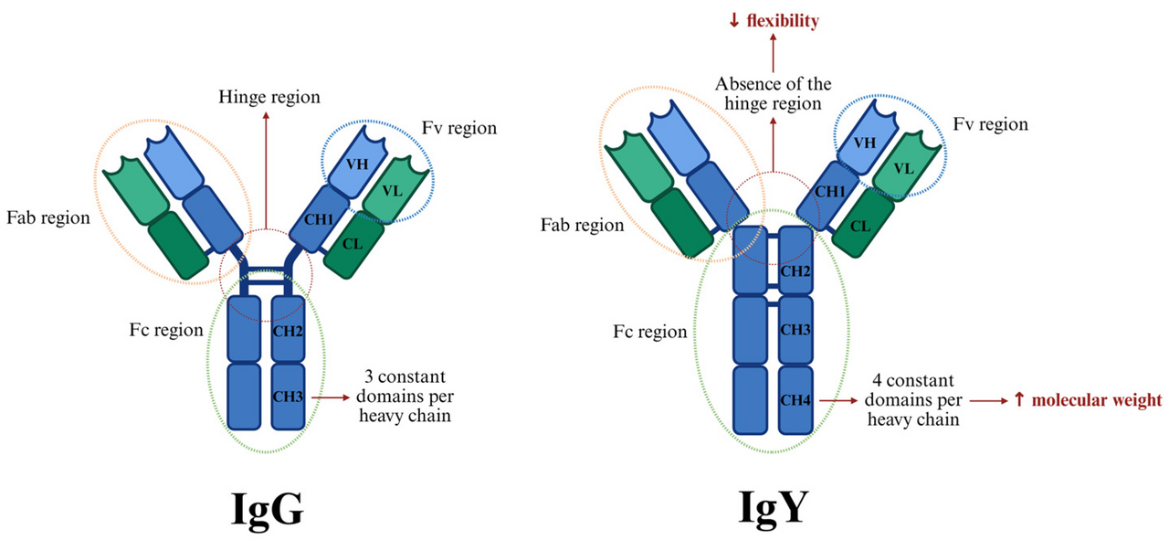
Birds do not possess IgD, and they have neither mammalian IgG nor IgE. Instead, IgY acts as the evolutionary progenitor of both. Structurally rigid and lacking the flexible, proline-rich hinge of mammalian IgG, IgY compensates with four heavy constant domains. It delivers systemic pathogen neutralization and complement activation in blood and tissues, while serving the exact function in eggs that mammalian IgG does across the placenta: providing the newborn chick with life-saving maternal immunity.
The Real-World Crucible: Vaccinating an Avian Immune System
This unusual evolutionary architecture explains why immunizing poultry–such as protecting commercial flocks against high-pathogenicity avian influenza (HPAI)–demands entirely different strategies than vaccinating humans or cattle:
- The Maternal IgY Moat: A newborn chick is saturated with maternal IgY absorbed straight from the yolk sac. Administering an inactivated flu vaccine during the first week or two is useless; the maternal antibodies immediately intercept and neutralize the vaccine antigen before the chick’s naive cells can see it.
- The Broiler Dilemma: Meat chickens (broilers) are bred to reach market weight in just 5 to 7 weeks. Because maternal antibodies take 2 to 3 weeks to wane to permissive levels, and an inactivated vaccine requires at least 2 weeks to induce protective titers, the bird’s operational window for conventional immunization is nearly nonexistent.
- The Viral Trojan Horse: To overcome both maternal antibody interference and rapid development timelines, veterinary medicine uses recombinant viral vectors, such as Herpesvirus of Turkeys (HVT) engineered to carry the influenza hemagglutinin (HA) gene. Injected in ovo directly into the egg at embryonic day 18 or at day of hatch, the cell-associated vector quietly establishes intracellular infection, producing the influenza target antigen from within host cells even under a sea of maternal yolk antibodies.
The avian immune system is neither primitive nor clumsy; it is a masterclass in streamlined biological engineering. By ditching the \(\kappa\) locus, running a short-lived copy-paste engine in the bursa, and equipping their circulation with ancestral IgY, birds solved the complex problem of adaptive immunity within the uncompromising constraints of flight.